|
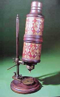
"Not Robert Hooke's own microscope as often claimed by the present owners ... but one very like it." |
As Robert Hooke (England) said in his book, Micrographia, a compound microscope is made from two magnifying glasses.
|
"Not Robert Hooke's own microscope as often claimed by the present owners ... but one very like it." |
The lower one (the objective) is of very short focal length. Some very early microscopes were made with a spherical glass bead for an objective lens. The object is placed just outside the focal length of the objective, forming a real enlarged image under the eyepiece, inside the focal length. The eyepiece acts like a regular magnifying glass.
The final virtual image is often formed at infinity, but in this case is shown at the near-point of the eye, so the diagram can be completed. [Many authors advise the formation of the final image at infinity to improve viewing comfort.] The angular magnification of the final image is affected very little by the final image position.
The magnification M is given at once by....
The final image is inverted. Moving an object to the right under a microscope moves the image to the left. People soon get used to this so there is no need to add correcting lenses.
Oil immersion microscopes, which dip the objective into a drop of oil on the coverslip which has the same refractive index as the lower lens, have improved fields of view and sharpened images but optical magnification is limited to about x1000 because of diffraction effects. A scanning UV microscope has a little more resolution at high magnifications but scanning electron microscopes, with amazing depth of field and resolution, are the ones used to make really spectacular micrographs.
Modern microscopes have more than two lenses.
Image quality is improved with a compound objective and the eyepiece has at least two lenses, one of which is an achromatic doublet.
Chromatic aberration occurs when rays of different colors (wavelengths) are not focused at the same place. The effect causes colored fringes to surround the image.
Chromatic aberration is eliminated [for two wavelengths only] by making lenses which are cemented doublets. The two lens components are made from different types of glass with different dispersion characteristics. Since perfect compensation is only possible for two wavelengths, chromatic aberration cannot be completely eliminated.
Note: the first microscopes made by Leeuwenhoek were single biconvex lens instruments. A replica is shown below.

The single lens was tiny, and had a very short focal length. The lens had to be placed very close to the eye. It appears that many of his contemporaries found Leeuwenhoek's instruments almost impossible to use, and doubted the authenticity of the drawings of what he claimed to see.
|
A word to the wise. The microscope ray diagram combines the two quite different diagrams for a convex lens and is a favorite with examiners. |